| Aug 22, 2024 |
Advancing nanoscale imaging capabilities
(Nanowerk News) Dynamic nuclear polarization (DNP) has revolutionized the field of nanoscale nuclear magnetic resonance (NMR), making it possible to study a wider range of materials, biomolecules and complex dynamic processes such as how proteins fold and change shape inside a cell.
|
|
A team of researchers at the University of Waterloo are combining pulsed DNP with nanoscale magnetic resonance force microscopy (MRFM) measurements to demonstrate that this process can be implemented on the nanoscale with high efficiency. The effort is overseen by Dr. Raffi Budakian, faculty member of the Institute for Quantum Computing and a professor in the Department of Physics and Astronomy, and his team consisting of Sahand Tabatabaei, Pritam Priyadarshi , Namanish Singh, Pardis Sahafi, and Dr. Daniel Tay.
|
|
The research has been published in Science Advances ("Large-Enhancement Nanoscale Dynamic Nuclear Polarization Near a Silicon Nanowire Surface").
|
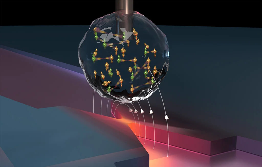 |
| Schematic of the experimental setup depicting a nanoscale droplet containing glucose and the free radical OX063. In the DNP process, the magnetic fields produced by the flow microwave frequency currents through a narrow metallic constriction induce coherent spin flips between electron spins in OX063 (green-coloured spheres) and nearby hydrogen spins (gold-coloured spheres). This exchange in magnetization boosts the polarization of hydrogen spins by more than a factor of 100, thus providing a significant enhancement in nuclear spin detection sensitivity. (Image: University of Waterloo)
|
|
In conventional magnetic resonance, the detection relies on the thermal population difference between “up” and “down” spin states within an external magnetic field. However, in nanoscale magnetic resonance, where the number of spins is significantly reduced, the inherent statistical fluctuations in spin orientation can be larger than the thermal polarization. Thus, it is better to measure the statistical polarization rather than the thermal polarization when observing nanoscale spin ensembles.
|
|
Nevertheless, due to the substantially larger thermal electron polarization compared to nuclear spins, dynamic nuclear polarization can be employed to amplify nuclear spin polarization by transferring polarization from electrons to nearby nuclei. This enhancement significantly boosts detection sensitivity in nuclear magnetic resonance applications.
|
|
The team’s experiments revealed a 100-fold increase in the thermal polarization of hydrogen nuclear spins, corresponding to a 15-fold increase in detection sensitivity, when compared to statistical polarization. Crucially, this enhancement corresponds to a reduction in the measurement time by a factor of 200, which allowed them to acquire signals much more rapidly. These results substantially advance the capabilities of MRFM detection as a practical tool for nanoscale imaging.
|
|
“By combining DNP's substantial enhancements with nanometer-scale magnetic resonance imaging (MRI) and ultra-sensitive spin detection, three-dimensional MRI of biomolecular structures with angstrom-scale resolution could become achievable — a transformative capability in structural biology,” Budakian says.
|
|
Looking forward, the research team aims to apply DNP-enhanced MRFM measurements for 3D nanometer scale structures such as viruses and proteins. They hope to increase nuclear spin detection sensitivity by operating at lower temperatures and higher magnetic fields.
|
