| Jul 05, 2024 |
Atomic force microscopy in 3D
(Nanowerk News) Atomic force microscopy (AFM) was originally invented for visualizing surfaces with nanoscale resolution. Its basic working principle is to move an ultrathin tip over a sample’s surface. During this xy-scanning motion, the tip’s position in the direction perpendicular to the xy-plane follows the sample’s height profile, resulting in a height map of the surface.
|
|
In recent years, ways to extend the method to three-dimensional (3D) imaging have been explored, with researchers from Nano Life Science Institute (WPI-NanoLSI), Kanazawa University reporting pioneering experiments on living cells. However, for 3D-AFM to evolve into a widely applicable technique for visualizing flexible molecular structures, a thorough understanding of the imaging mechanisms at play is necessary.
|
|
Now, Takeshi Fukuma from Kanazawa University and colleagues have performed a detailed study of a specially designed flexible sample, providing essential insights into the theoretical basis and the interpretation of 3D-AFM experiments.
|
|
They report their findings in Small Methods ("Revealing the Mechanism Underlying 3D-AFM Imaging of Suspended Structures by Experiments and Simulations").
|
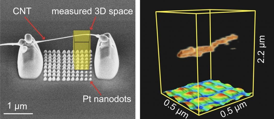 |
| Imaged nanostructure consisting of a suspended carbon nanotube with platinum nanodots beneath. Left: 3D model of vertical nanostructures. Right: Three-dimensional atomic force microscopy (3D-AFM) map underlining its capability to image suspended flexible samples above a regular pattern of nano-sized dots. (Image: Mohammad Shahidul Alam, et al.)
|
|
Using microfabrication tools, the scientists created a sample consisting of a carbon nanotube fiber resting on platinum pillars that in turn were positioned on a silicon substrate. A carbon nanotube is a structure that one can think of as a rolled-up, one-atom-thick carbon sheet. The freestanding portion of the nanotube was about 2 micrometers long. The whole structure was immersed in water, as many 3D biomolecular systems of interest occur in liquid environments.
|
|
Fukuma and colleagues then performed 3D-AFM experiments in two different modes. In static mode, the nanotip is lowered vertically towards the sample. When the tip makes contact with the suspended nanotube fiber, the latter gets pushed aside, and bends while the probe descends further. In dynamic mode, the tip, which is attached to a cantilever, is made to oscillate at a resonance frequency while being lowered.
|
|
By analyzing how the force experienced by the tip changes as a function of the tip’s depth, the researchers concluded that the friction between the tip and the fiber is much larger in static mode compared to dynamic mode. The latter is therefore the mode of choice, as less friction means that potential damage to the sample is less likely.
|
|
The scientists performed computer simulations to model what happens when the tip reaches the carbon nanotube fiber. The simulations confirmed that the suspended nanotube displaces laterally, and that a continuously vibrating tip (as in dynamical mode) results in weaker forces experienced by the sample, hindering strong adhesion of the tip to the fiber.
|
|
Fukuma and colleagues then performed experiments with a carbon nanotube fiber suspended above a regular pattern of nano-sized platinum dots deposited on a silicon substrate. The measurements were done in dynamical mode. The reconstructed 3D map of the scanned volume clearly showed the fiber and the dots below it, underlining the capability of 3D-AFM to image vertically overlapping nanostructures.
|
|
These findings show that AFM can generally be applied to visualize flexible 3D structures. Quoting the scientists: “… the advancements made in this study may potentially lead to more detailed and accurate AFM analysis of various 3D biological systems such as cells, organelles, chromosomes, and vesicles.”
|
Background
|
|
The principle behind atomic force microscopy (AFM) is to scan the surface of a sample with a very small tip. During this horizontal (xy) scan, the tip, attached to a small cantilever, follows the sample’s vertical (z) profile, which induces a force on the cantilever that can be measured. The magnitude of the force at the xy position can be related to the z value. The xyz data generated during a scan then result in a height map providing structural information about the investigated sample. The cantilever can be made to oscillate near its resonance frequency, which is referred to as dynamic mode AFM. Not letting the cantilever oscillate is known as static mode AFM. In dynamic mode, when the tip is moved around a surface, the variations in the amplitude (or the frequency) of the cantilever’s oscillation — resulting from the tip’s interaction with the sample’s surface — are recorded, as these provide a measure for the local z value.
|
|
Takeshi Fukuma and colleagues have now provided a detailed AFM analysis of a 3D reference sample with nanosized features that could be reconstructed with high precision. The experiments and accompanying simulations confirm that AFM has the potential to become a robust method for the characterization of 3D nanosized objects, including biological systems.
|
