Scientists develop 'X-ray vision' technique to see inside crystals
(Nanowerk News) A team of New York University researchers has created a new way to visualize crystals by peering inside their structures, akin to having X-ray vision. Their new technique – which they aptly named “Crystal Clear” – combines the use of transparent particles and microscopes with lasers that allow scientists to see each unit that makes up the crystal and to create dynamic three-dimensional models.
“This is a powerful platform for studying crystals,” says Stefano Sacanna, professor of chemistry at NYU and the principal investigator for the study, published in the journal Nature Materials ("Enabling three-dimensional real-space analysis of ionic colloidal crystallization"). “Previously, if you looked at a colloidal crystal through a microscope, you could only get a sense of its shape and structure of the surface. But we can now see inside and know the position of every unit in the structure.”
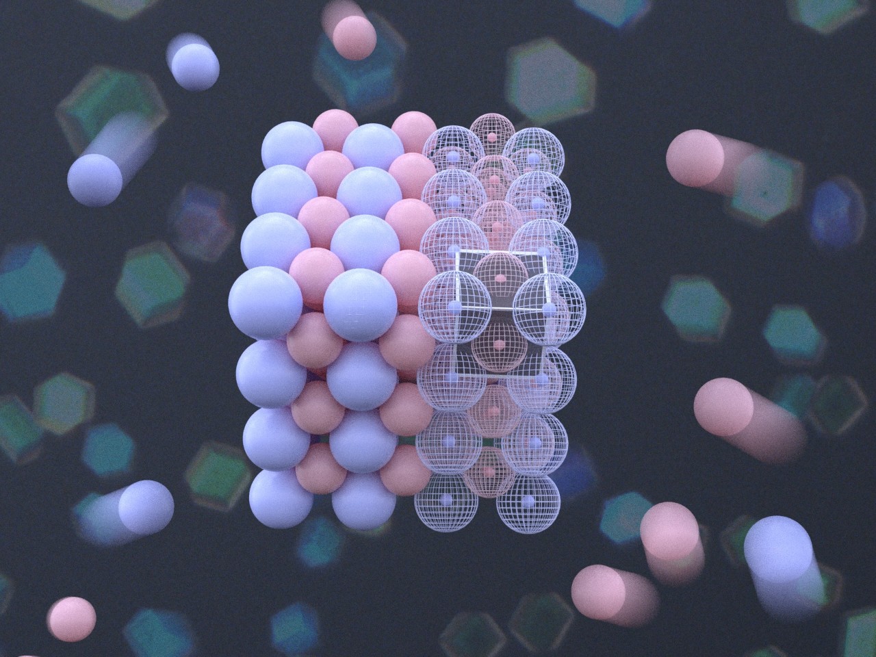
The new technique allows scientists to see each particle that makes up colloidal crystals and to create dynamic three-dimensional models. (Image: Shihao Zang)
Atomic crystals are solid materials whose building blocks are positioned in a repeating, orderly fashion. Every now and then, an atom is missing or out of place, resulting in a defect. The arrangement of atoms and defects is what creates different crystalline materials—from table salt to diamonds—and gives them their properties.
To study crystals, many scientists, including Sacanna, look to crystals composed of miniscule spheres called colloidal particles rather than atoms. Colloidal particles are tiny – often around a micrometer in diameter, or dozens of times smaller than a human hair – but are much larger than atoms and therefore easier to see under a microscope.
A see-through structure
In their ongoing work to understand how colloidal crystals form, the researchers recognized the need to see inside these structures. Led by Shihao Zang, a PhD student in Sacanna’s lab and the study’s first author, the team set out to create a method to visualize the building blocks inside a crystal. They first developed colloidal particles that were transparent and added dye molecules to label them, making each particle possible to distinguish under a microscope using their fluorescence.
A microscope alone wouldn’t allow the researchers to see inside a crystal, so they turned to an imaging technique called confocal microscopy, which uses a laser beam that scans through material to produce targeted fluorescence from the dye molecules. This reveals each two-dimensional plane of a crystal, which can be stacked on top of each other to build a three-dimensional digital model and identify the location of each particle. The models can be rotated, sliced, and taken apart to look inside the crystals and see any defects.
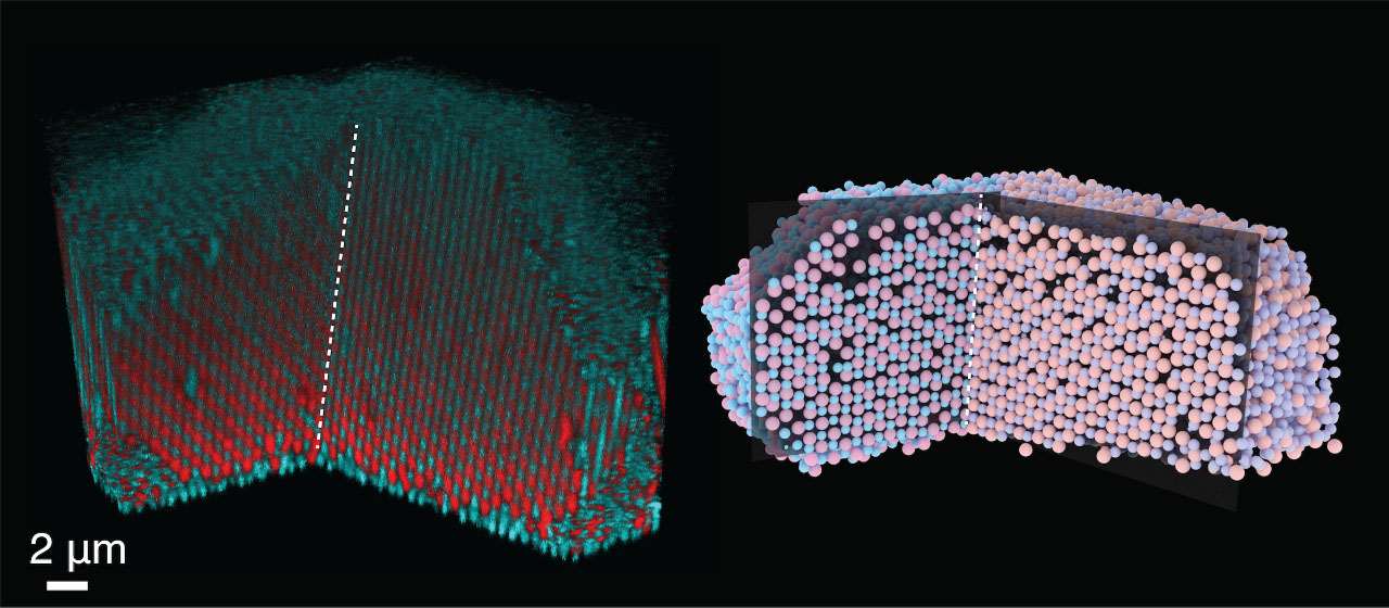
In one set of experiments, the researchers used this imaging method on crystals that form when two of the same type of crystals grow together—a phenomenon known as “twinning.” When they looked inside models of crystals having structures equivalent to table salt or an alloy of copper and gold, they could see the shared plane of the adjoined crystals, a defect that gives rise to these particular shapes. This shared plane revealed the molecular origin of twinning.
A 3D scan and digital model of crystal "twinning" reveals a shared plane of the adjoining crystals, which gives rise to their shape. (Image: Shihao Zang)
Crystals in motion
In addition to looking at static crystals, this new technique allows scientists to visualize crystals as they change. For example, what happens when crystals melt—do particles rearrange, and do defects move? In an experiment in which the researchers melted a crystal with the structure of the mineral salt cesium chloride, they were surprised to find that the defects were stable and did not move around as expected.
In order to validate their experiments on static and dynamic crystals, the team also used computer simulations to create crystals with the same characteristics, confirming that their “Crystal Clear” method accurately captured what is inside crystals.
“In a sense, we’re trying to put our own simulations out of business with this experiment—if you can see inside the crystal, you may not need simulations anymore,” jokes Glen Hocky, assistant professor of chemistry at NYU, a faculty member in the Simons Center for Computational Physical Chemistry at NYU, and the study’s co-corresponding author.
Now that scientists have a method for visualizing the inside of crystals, they can more easily study their chemical history and how they form, which could pave the way for building better crystals and developing photonic materials that interact with light.
“Being able to see inside crystals gives us greater insight into how the crystallization process works and can perhaps help us to optimize the process of growing crystals by design,” adds Sacanna.
Source: By Rachel Harrison, NYU (Note: Content may be edited for style and length)
We curated lists with the (what we think) best science and technology podcasts - check them out!