| Dec 30, 2020 |
New X-ray camera achieves new heights of precision and accuracy
(Nanowerk News) Scientists use incredibly bright and fast pulses of X-rays produced by an X-ray free electron laser to study some of the fastest reactions and processes in materials. X-rays in these experiments can have wavelengths of less than an Angstrom—more than one million times smaller than the diameter of a human hair.
|
|
Scientists recently developed a new X-ray imager with much greater precision and accuracy than possible before. The new levels are less than one hundredth of an X-ray wavelength, even smaller than an Angstrom. These X-ray imagers are becoming a useful tool in research that uses X-ray free electron lasers.
|
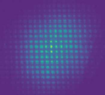 |
| An X-ray image taken with a novel X-ray wavefront imager results in high precision measurements of intensity and direction of the X-ray beam. (Image: SLAC National Accelerator Laboratory)
|
|
In an X-ray free electron laser experiment, researchers must know the properties of the X-rays as they come out of the laser, go through instruments, and interact with the material being studied.
|
|
In this new research, scientists used measure the intensity distribution of the X-rays going into the sample, but also determine the directions at which these X-rays are traveling. Knowing these X-ray properties can improve experimental analysis.
|
|
Imaging materials with a high-frequency X-ray free electron laser pulses can be challenging because the X-rays coming from the laser can have fluctuations in their properties from pulse to pulse.
|
|
The ability to image the X-ray wavefront a single shot at a time is important not only for experimental design and analysis, but also as feedback to allow laser and instrument optimization. |
|
This research developed a single-shot wavefront imager composed of a single 2D grating and a scintillator-based detector. It uses the Talbot effect, an effect where the X-rays are “self-imaged” as they travel.
|
|
The Talbot effect is generated using a 2D grating made with nanofabrication techniques in either diamond or silicon, engineered to have very low distortion. By recording the “self-image” after the grating, both the amplitude and phase of the X-rays can be determined using a reconstruction algorithm.
|
|
The simplicity of the wavefront imager makes it easier to place the imager at different parts of the beam path in an experiment. For example, researchers could place the imager at the exit of the free electron laser to provide real-time feedback.
|
|
Similarly, the imager could be used downstream of various components of the instrument to provide systemic understanding and feedback as the X-rays travel or to directly image materials. Researchers have demonstrated this wavefront imager over a wide range of X-ray energies with extremely high accuracy and precision.
|
Publications
|
|
Liu, Y. et al., X-ray free-electron laser wavefront sensing using the fractional Talbot effect. J. Synchrotron Radiation 27, 254-261 (2020). [DOI: 10.1107/S1600577519017107]
|
|
Li, K. et al., Wavefront preserving and high efficiency diamond grating beam splitter for x-ray free electron laser. Optics Express 28, 10939-10950 (2020). [DOI: 10.1364/OE.380534]
|
|
Liu, K. et al. High-accuracy wavefront sensing for x-ray free electron lasers. Optica 5, 967-975 (2018). [DOI: 10.1364/OPTICA.5.000967]
|
