Tip-Enhanced Raman Spectroscopy (TERS): Nanoscale Chemical Analysis at the Tip of a Needle
What is Tip-Enhanced Raman Spectroscopy?
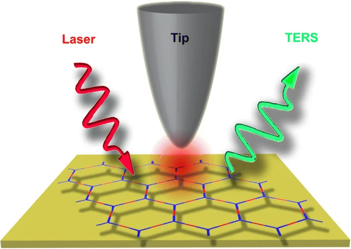
Tip-enhanced Raman spectroscopy (TERS) is a powerful analytical technique that combines the high spatial resolution of scanning probe microscopy (SPM) with the molecular specificity of Raman spectroscopy. It enables the acquisition of chemical and structural information at the nanoscale, far beyond the diffraction limit of conventional optical microscopy.
Key Concepts in TERS
TERS is based on several key concepts that enable its exceptional performance:
- Surface Plasmon Enhancement: TERS exploits the localized surface plasmon resonance (LSPR) effect, which occurs when a metallic nanostructure, such as a sharp tip, is illuminated with light. The LSPR leads to a strong enhancement of the electromagnetic field at the tip apex, which significantly amplifies the Raman scattering from nearby molecules.
- Near-Field Excitation: In TERS, the tip is brought into close proximity (a few nanometers) to the sample surface, allowing the enhanced electromagnetic field to interact with the molecules in the near-field region. This near-field interaction enables the detection of Raman signals from extremely small volumes, typically on the order of a few cubic nanometers.
- High Spatial Resolution: The spatial resolution of TERS is determined by the size and shape of the tip apex, rather than the diffraction limit of light. With carefully designed tips, TERS can achieve spatial resolutions down to a few nanometers, enabling the investigation of individual molecules, nanostructures, and even atomic-scale features.
Instrumentation and Methodology
TERS requires a specialized instrumentation setup that integrates an SPM system with a Raman spectrometer:
SPM System
The SPM system, typically an atomic force microscope (AFM) or a scanning tunneling microscope (STM), is used to position and control the tip with nanoscale precision. The tip is usually made of gold or silver and is chemically etched or electrochemically polished to obtain a sharp apex with a radius of curvature of a few nanometers.
Raman Spectrometer
The Raman spectrometer is used to excite the sample with a focused laser beam and collect the scattered light. The laser wavelength is chosen to match the LSPR of the tip, maximizing the field enhancement. The scattered light is then dispersed by a grating and detected by a sensitive CCD camera or a single-photon counting module.
TERS Modes
TERS can be operated in different modes, depending on the sample and the desired information:
- Point Spectroscopy: The tip is positioned at a specific location on the sample, and a Raman spectrum is acquired. This mode provides detailed chemical information at a single point.
- Line Scanning: The tip is scanned along a line on the sample surface, and Raman spectra are collected at regular intervals. This mode generates a one-dimensional chemical map of the sample.
- Raster Scanning: The tip is raster-scanned over a two-dimensional area on the sample, and Raman spectra are acquired at each pixel. This mode produces a high-resolution chemical image of the sample surface.
Applications of TERS
TERS has found applications in various fields, including materials science, nanoscience, and life sciences:
Materials Characterization
TERS is widely used for the characterization of nanomaterials, such as graphene, carbon nanotubes, and semiconductor nanostructures. It provides valuable insights into their local chemical composition, defects, strain, and electronic properties.
Surface and Interface Analysis
TERS is a powerful tool for studying surfaces and interfaces at the nanoscale. It can probe the chemical composition and molecular orientation of thin films, self-assembled monolayers, and heterogeneous catalysts, providing a deeper understanding of surface phenomena and reactions.
Biological and Biomedical Applications
TERS has emerged as a promising technique for the investigation of biological systems at the nanoscale. It has been applied to study the chemical composition of cell membranes, protein structures, and DNA, offering new insights into the molecular basis of biological processes and diseases.
Challenges and Future Perspectives
Despite the remarkable capabilities of TERS, several challenges need to be addressed for its widespread adoption. One of the main challenges is the reproducibility and stability of the TERS tips. The fabrication of high-quality tips with consistent enhancement factors and long lifetimes remains a critical issue.
Future research in TERS will focus on the development of novel tip materials and geometries to improve the signal enhancement and spatial resolution. The integration of TERS with other advanced spectroscopic and imaging techniques, such as super-resolution microscopy and tip-enhanced coherent anti-Stokes Raman scattering (TE-CARS), will further expand its capabilities. Additionally, the application of machine learning algorithms for data analysis and interpretation will facilitate the extraction of meaningful chemical information from complex TERS datasets.
Further Reading
EPJ Techniques and Instrumentation, Tip-enhanced Raman spectroscopy: principles and applications
Chemical Reviews, Tip-Enhanced Raman Spectroscopy: Technique and Recent Advances
Chemical Society Reviews, Tip-enhanced Raman spectroscopy for surfaces and interfaces
