Scanning X-Ray Fluorescence (XRF) Microscopy: Unveiling the Elemental Composition of Nanomaterials
What is Scanning X-Ray Fluorescence (XRF) Microscopy?
Scanning X-Ray Fluorescence (XRF) Microscopy is a powerful analytical technique that enables the visualization and quantification of the elemental composition of materials at the nanoscale. It combines the principles of X-ray fluorescence spectroscopy with high-resolution scanning microscopy to provide spatially resolved elemental maps of samples.
Historical Context and Development
The development of Scanning X-Ray Fluorescence (XRF) Microscopy can be traced back to the early days of X-ray fluorescence spectroscopy in the early 20th century. The technique evolved from bulk XRF analysis to spatially resolved measurements with the advent of focused X-ray beams and scanning stages.
Key milestones in the development of Scanning XRF Microscopy include:
- 1950s-1960s: The introduction of the first X-ray microprobes, which used collimated X-ray beams to analyze small sample areas.
- 1980s: The development of synchrotron radiation sources, which provided high-intensity, tunable X-rays for improved spatial resolution and sensitivity.
- 1990s-2000s: Advancements in X-ray optics, such as Kirkpatrick-Baez mirrors and zone plates, enabled the focusing of X-ray beams to submicron and nanometer sizes, leading to the emergence of high-resolution scanning XRF microscopy.
- 2000s-Present: Continuous improvements in detector technology, data acquisition, and analysis methods have further enhanced the capabilities and applications of scanning XRF microscopy in various fields, including materials science, biology, and environmental studies.
Today, scanning XRF microscopy is a well-established and widely used technique for the elemental characterization of nanomaterials and other samples, offering unique insights into the spatial distribution and quantification of elements at the nanoscale.
Principles of Scanning XRF Microscopy
Scanning XRF Microscopy is based on the following key principles:
- X-Ray Fluorescence: When a sample is irradiated with high-energy X-rays, the atoms in the sample absorb the X-rays and become ionized. As the excited electrons return to their ground state, they emit characteristic X-rays with energies specific to each element.
- Scanning Microscopy: A focused X-ray beam is scanned across the sample surface in a raster pattern. At each point, the emitted X-rays are detected and analyzed to determine the elemental composition at that specific location.
- Elemental Mapping: By correlating the X-ray fluorescence data with the scanning position, elemental maps can be generated, providing a visual representation of the spatial distribution of elements within the sample.
Instrumentation
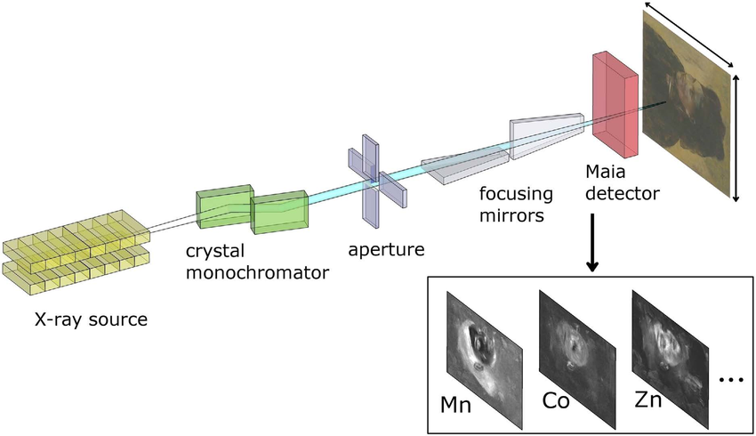
A typical scanning XRF microscope consists of the following components:
- X-Ray Source: A high-intensity X-ray source, such as a synchrotron or a microfocus X-ray tube, is used to generate the incident X-rays. The X-ray beam is focused onto the sample using X-ray optics, such as Kirkpatrick-Baez mirrors or zone plates.
- Sample Stage: The sample is mounted on a motorized stage that allows precise positioning and scanning of the sample relative to the X-ray beam.
- X-Ray Detector: An energy-dispersive X-ray detector, such as a silicon drift detector (SDD) or a silicon lithium (Si(Li)) detector, is used to collect the emitted X-rays and measure their energies.
- Data Acquisition and Analysis: The X-ray fluorescence data is acquired and processed using specialized software that generates elemental maps, quantifies elemental concentrations, and performs data analysis.
Capabilities and Applications
Scanning XRF Microscopy offers several unique capabilities and finds applications in various fields:
- High Spatial Resolution: Scanning XRF microscopes can achieve spatial resolutions down to tens of nanometers, enabling the study of elemental distributions at the nanoscale.
- Quantitative Analysis: By calibrating the X-ray fluorescence signal against standards, quantitative information about elemental concentrations can be obtained.
- Non-Destructive Analysis: Scanning XRF microscopy is a non-destructive technique, allowing the analysis of valuable or irreplaceable samples without causing damage.
- Material Characterization: Scanning XRF microscopy is widely used for the characterization of nanomaterials, including nanoparticles, nanowires, and thin films. It provides insights into the elemental composition, homogeneity, and spatial distribution of elements within the sample.
- Biological and Environmental Applications: Scanning XRF microscopy is applied in the study of biological samples, such as cells and tissues, to investigate the distribution and role of trace elements. It is also used in environmental science to analyze the elemental composition of particulate matter, sediments, and other environmental samples.
- Cultural Heritage and Art Conservation: Scanning XRF microscopy is employed in the analysis of historical artifacts, paintings, and manuscripts to study the materials used, the presence of contaminants, and the conservation state of the objects.
Limitations and Challenges
While scanning XRF microscopy is a powerful technique, it also has some limitations and challenges:
- Sample Preparation: Samples need to be prepared in a way that is compatible with the X-ray beam and the scanning stage. This may require thin sectioning, embedding, or other sample preparation techniques.
- Matrix Effects: The X-ray fluorescence signal can be influenced by the matrix in which the elements are present. Matrix effects need to be considered and corrected for accurate quantification.
- Overlapping X-Ray Lines: Some elements have overlapping X-ray fluorescence lines, which can complicate the analysis and require careful deconvolution of the spectra.
- Synchrotron Access: High-resolution scanning XRF microscopy often requires access to synchrotron facilities, which can be limited and competitive.
Future Perspectives
Scanning XRF Microscopy continues to evolve and improve, with ongoing developments in instrumentation, data analysis, and applications. Some future perspectives include:
- Improved Spatial Resolution: Advancements in X-ray optics and detectors are expected to push the spatial resolution of scanning XRF microscopy even further, enabling the study of nanomaterials at ever-smaller scales.
- In Situ and Operando Studies: The development of specialized sample environments and stages will allow in situ and operando studies, where the elemental composition can be monitored during dynamic processes or under various environmental conditions.
- Integration with Complementary Techniques: Combining scanning XRF microscopy with other techniques, such as X-ray diffraction, X-ray absorption spectroscopy, or Raman spectroscopy, will provide a more comprehensive understanding of the sample's structural, chemical, and functional properties.
Further Reading
Current Opinion in Chemical Biology, Biological applications of X-ray fluorescence microscopy: exploring the subcellular topography and speciation of transition metals
