Showing Spotlights 297 - 304 of 544 in category All (newest first):
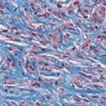
 Candida albicans is a leading fungal cause of burn infections in hospital settings. The prevalence of invasive candidiasis in burn cases varies widely, but it accounts as high as 23% of severe infection with a mortality rate ranging from anywhere to 14% to 70%. In a recent pre-clinical study, a nitric oxide releasing nanoparticle platform, which has previously been shown to be antibacterial to both gram positive and negative bacteria, as well as an accelerator of wound healing in excisional animal models, was found to be efficacious in clearing candidal burn infections in mice. This study represents one of many pre-clinical investigations demonstrating the efficacy of the NO nanoparticles as a broad spectrum antimicrobial agent as well as wound healing accelerant.
May 21st, 2012
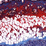
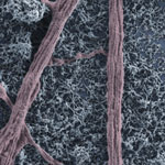
 Wound healing is an exceedingly complex process, involving a multitude of signaling pathways, effector molecules, response phases, as well as a moderated balance between all these components. Nitric oxide (NO) plays a critical role in the wound-healing process via antimicrobial properties, modulation of platelet/cytokine function, vasodilatory effects, and promotion of angiogenesis and matrix deposition. While attempts to administer NO to wound areas have shown some promise, the current modalities all suffer from varying drawbacks, such as administration site irritation or the burden of large, expensive equipment. Researchers have now introduced a nanoparticle platform comprised of silane based sol-gel and sugar-derived glasses that can generate, store, and deliver NO in a controlled and sustained manner is utilized to enhance wound healing in immunodeficient mice.
Mar 28th, 2012
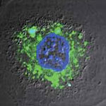
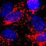 Nanotechnology offers new strategies to enable minimally invasive and localized approaches for diagnosing and treating cancer, thereby avoiding the serious side effects and shortcomings of chemotherapy. For instance, it has been shown that often less than 1% of the administered drug molecules during chemotherapy enter tumor cells and bind to the nuclear DNA. Another complication is drug resistance of cancer cells. This actually is one of the main causes of failure in the treatment of cancer. Cancer researchers are looking to nanoparticles as a drug carrier capable of localizing and directly releasing drugs into the cell nucleus, leading to a high therapeutic efficacy. Although increased therapeutic efficacy has been realized, there have been no reports on visualizing at nanoscale dimensions how nanoparticles interact with specific organelles. In a new breakthrough for nanomedicine cancer research, scientists have now reported the direct visualization of interactions between drug-loaded nanoparticles and the nucleus of a cancer cell.
Mar 26th, 2012
 Green Fluorescent Protein (GFP) - originally found in a jellyfish - has played a crucial role in life science research, providing insights to many fundamental questions that have paved the way to the biology and medicine of the future. Since the mid-1990s, when the protein was successfully cloned, GFP can be found in research laboratories worldwide used as a visual marker of gene expression and protein localization, easily observed via light (optical) microscopy. GFP can be linked to other proteins and is primarily used to track dynamic changes in living cells. In 2008, biologists who discovered and developed the protein as a laboratory tool won a Nobel Prize for their work. Researchers in Spain have now demonstrated how GFP can also act as an efficient nano-thermometer inside cells.
Mar 19th, 2012
 Carbon nanotubes, like the nervous cells of our brain, are excellent electrical signal conductors and can form intimate mechanical contacts with cellular membranes, thereby establishing a functional link to neuronal structures. There is a growing body of research on using nanomaterials in neural engineering. Now, researchers have, for the first time, explored the impact of carbon nanotube scaffolds on multilayered neuronal networks. Up to now, all known effects of carbon nanotubes on neurons - namely their reported ability to potentiate neuronal signaling and synapses - have been described in bi-dimensional cultured networks where nanotube/neuron hybrids were developed on a monolayer of dissociated brain cells.
Mar 2nd, 2012
 Carbon nanotubes (CNTs) offer a number of advantages for delivering drugs to specific locations inside the body which suggest that they may provide an improved result over nanoparticles. They have a larger inner volume which allows more drug molecules to be encapsulated, and this volume is more easily accessible because the end caps can be easily removed, and they have distinct inner and outer surfaces for functionalization. Recent research has shown the ability of CNTs to carry a variety of molecules such as drugs, DNA, proteins, peptides, targeting ligands etc. into cells - which makes them suitable candidates for targeted delivery applications. Researchers have now developed a unique two-dye labeling method to directly track the release process of a anti-cancer drug from carbon nanotube carriers in living cells.
Mar 1st, 2012
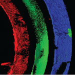 At the core of tissue engineering is the construction of three-dimensional scaffolds out of biomaterials to provide mechanical support and guide cell growth into new tissues or organs. In another advance for the field, researchers have now demonstrated a strategy to fabricate tubular structures with multiple types of cells as different layers of the tube walls. This method may be widely used in simulation of many tubular tissues and enriches the toolbox for 3D micro/nanofabrication by initially patterning in 2D and transforming it into 3D. Tubular tissues such as the trachea, blood vessels, lymph vessels, and intestines, have two distinguishing features: They have specific 3D shapes;and they have different types of cells at specific locations, i.e. different parts of the tube wall are made up of different cells. Mimicking both of these features is a prerequisite for fabricating functional tubular tissues in vitro, and the realization of structural-tissue mimicry may have wide applications.
Feb 14th, 2012
 With the advent of nanomedicine, the concept of a "magic bullet" to fight cancer is getting closer to reality. Previously an idea straight out of science fiction, researchers around the world are working on perfecting nano- and microscale drug carriers that get injected into the body, transport themselves to the correct target, such as a tumor, and deliver the required dose of a medication or other substance to effectively destroy or repair this target. The controlled drug release required by these systems, however, has proven to be quite a challenging issue. To avoid the side effects of prematurely released toxic cancer drugs on healthy tissues, researchers have designed and fabricated an "active defense" system which could effectively keep the drug entrapped in its carrier in the blood and normal tissues whereas it would allow the explosive drug release under the right physiopathological stimuli once the drug carrier reaches the cancerous tissues.
Feb 13th, 2012
 Subscribe to our Nanotechnology Spotlight feed